The Role of Cell Block in Diagnosing Metastatic Adenocarcinoma in Pleural Fluid: A Case Series
Article Sidebar
Main Article Content
Abstract
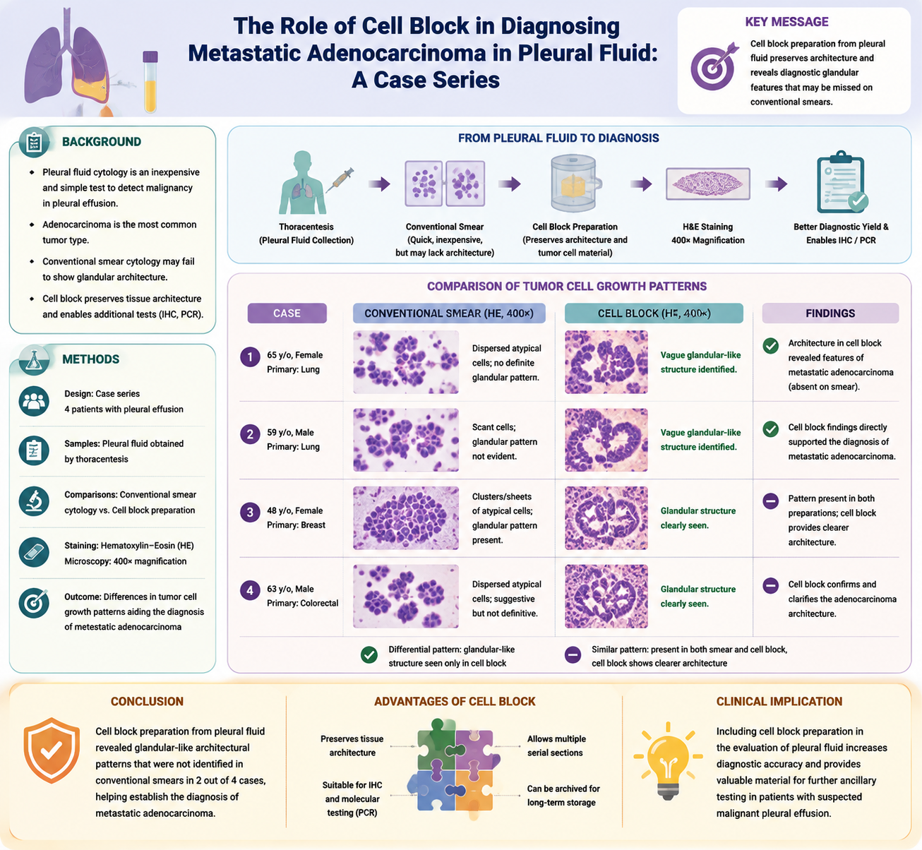
Background: Cytological examination of pleural fluid is an important, inexpensive, and simple test for detecting malignancy in patients with clinical pleural effusion who undergo thoracentesis. Adenocarcinoma is the most common tumor type. However, diagnosis using the conventional smear method can be challenging when the typical glandular-like growth patterns are not visible. Cell block preparation from pleural fluid samples preserves tissue architecture and tumor cell genetic material, enabling further tests such as immunohistochemistry (IHC) and PCR.
Case presentation: We report a series of four patients with pleural effusion who underwent both conventional smear cytology and cell block examination. Tumor cell growth patterns were identified and descriptively analyzed for each sample. In two of these cases, vague glandular-like growth patterns were observed only in the cell block preparations and were absent in the conventional smears, which directly helped establish the diagnosis of metastatic adenocarcinoma.
Conclusion: Differences in tumor cell growth patterns between the conventional smears and the cell block preparations were observed in two cases. The vague glandular-like growth pattern that was present in the cell block but was not identified in the conventional smear aided pathologists in identifying metastatic adenocarcinoma in pleural fluid specimens.
Article Details

This work is licensed under a Creative Commons Attribution 4.0 International License.
How to Cite
References
Geçmen GG. Cytology of pleural effusions. South Clin Ist Euras. 2025;36(1):35-41.
Ugurluoglu C, Kurtipek E, Unlu Y, Esme H, Duzgun N. Importance of the cell block technique in diagnosing patients with non-small cell carcinoma accompanied by pleural effusion. Asian Pac J Cancer Prev. 2015;16(7):3057-60.
Mathew JK, Rajan GN, Kunju AK. Diagnostic analysis of pleural fluid cell blocks using relevant immunohistochemical markers in clinically suspicious cases of malignancy. CytoJournal. 2024;21:8.
Porcel JM, Quirós M, Gatius S, Bielsa S. Examination of cytological smears and cell blocks of pleural fluid: complementary diagnostic value for malignant effusions. Rev Clin Esp (Barc). 2017;217(3):144-8.
Rajeev R, Suma M, Valooran JL. Cytological diagnosis of malignant pleural effusion caused by metastatic squamous cell carcinoma: a series of four cases. J Clin Diagn Res. 2024;18(1):ED01-3.
Rachmadi L, Setyoheriyanto D, Kusumastuti EH, Agustina H, Sembiring RE, Bilianti YD. Penanganan spesimen, diagnosis, imunohistokimia dan patologi molekular pada karsinoma paru. Jakarta: Perhimpunan Dokter Spesialis Patologi Anatomi Indonesia; 2019.
Torous VF, Cuda JM, Manucha V, Randolph ML, Shi Q, VandenBussche CJ. Cell blocks in cytology: review of preparation methods, advantages, and limitations. J Am Soc Cytopathol. 2023;12(2):77-88.
Assawasaksakul T, Boonsarngsuk V, Incharoen P. A comparative study of conventional cytology and cell block method in the diagnosis of pleural effusion. J Thorac Dis. 2017;9(9):3161-7.
Wang W, Tang Y, Li J, Jiang L, Jiang Y, Su X. Detection of ALK rearrangements in malignant pleural effusion cell blocks from patients with advanced non-small cell lung cancer: a comparison of Ventana immunohistochemistry and fluorescence in situ hybridization. Cancer Cytopathol. 2015;123(2):117-22.
Wang H, Agulnik J, Kasymjanova G, Wang A, Jiménez P, Cohen V, et al. Cytology cell blocks are suitable for immunohistochemical testing for PD-L1 in lung cancer. Ann Oncol. 2018;29(6):1417-22.
Zou Y, Xu L, Tang Q, You Q, Wang X, Ding W, et al. Cytology cell blocks from malignant pleural effusion are good candidates for PD-L1 detection in advanced NSCLC compared with matched histology samples. BMC Cancer. 2020;20(1):344.
Wang W, Tang Y, Li J, Zhang Y, Zou Y, Su X. Identification of EGFR mutations in cytological specimens of non-small cell lung carcinoma from a single institute. Int J Clin Exp Pathol. 2018;11(2):929-35.
Man Ng JK, Chow C, Kin Chan RC, Pang Chan K, Xi Li JJ, Ching Li MS, et al. EGFR testing in paraffin-embedded cell block cytology material is reliable with increased detection for effusion fluid. Lung Cancer. 2022;174:97-103.
Ranieri SC, Di Leonardo G, Coletti G, Dal Mas A, Brancone ML, Crisci R, et al. Role of conventional cytology and cell block methods for diagnosis of malignant pleural effusions. J Xiangya Med. 2020;5:36.
Rani SSS, Vamshidhar IS, John NA, John J. Diagnosis of pleural effusions by cell block and pleural biopsy: a comparative study. J Cytol. 2022;39(4):169-73.
Shivakumarswamy U, Arakeri SU, Karigowdar MH, Yelikar B. Diagnostic utility of the cell block method versus the conventional smear study in pleural fluid cytology. J Cytol. 2012;29(1):11-5.
Kumar SH, S S, Shetty D, Rao R. Clinicopathological study of 117 body fluids: comparison of conventional smear and cell block technique. Curr Health Sci J. 2020;46(4):336-43.
Nindrea RD, Aryandono T, Lazuardi L, Dwiprahasto I. Protective Effect of Omega-3 Fatty Acids in Fish Consumption Against Breast Cancer in Asian Patients: A Meta-Analysis. Asian Pac J Cancer Prev. 2019;20(2):327-32.
Thapar M, Mishra RK, Sharma A, Goyal V, Goyal V. Critical analysis of cell block versus smear examination in effusions. J Cytol. 2009;26(2):60-4.
Dey S, Nag D, Nandi A, Bandyopadhyay R. Utility of cell block to detect malignancy in fluid cytology: adjunct or necessity? J Cancer Res Ther. 2017;13(3):425-9.